Lean tissue on a body scan includes more than muscle
By Mr.Apps · Nov 2, 2026
Category:Energy

What lean tissue on a scan includes
Lean tissue on a body scan includes more than muscle. A dual-energy X-ray absorptiometry scan, often called DXA, separates the body into reported compartments such as fat mass, lean soft tissue, and bone mineral content. The scan estimates these components from how the measured signals pass through different tissues. It does not isolate each skeletal muscle and weigh it directly.
That distinction matters when a report is used to review training progress, nutrition, or a change in health. Lean soft tissue includes skeletal muscle, but it can also include water, organs, skin, connective tissue, and other nonfat soft tissue. A shift in the reported compartment is not automatically the same as a change in contractile muscle. The scan is useful when its method, region, and limitations stay attached to the result.
The body-composition terminology guide distinguishes lean soft tissue from skeletal muscle and explains the compartments DXA reports. The language on a device or clinic report may be shorter or less precise than the scientific term. I keep the exact label used by the report before interpreting what a change might mean.
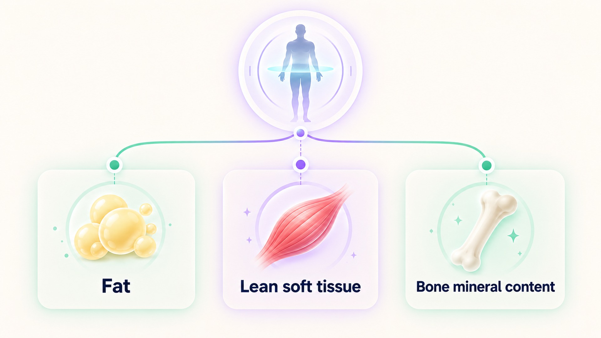
How the scan divides body mass
A DXA scanner estimates fat mass, lean soft tissue, and bone mineral content. Those categories describe different components. Lean soft tissue refers to soft tissue after the fat and bone mineral components are separated according to the method. It is not a direct measure of one tissue type. In a body region that contains bone, the scan's classification also depends on how the image-processing method distinguishes the bone mineral from surrounding tissue.
Skeletal muscle is an organ-level tissue made of muscle fibers and associated structures. Lean soft tissue is a broader compartment reported from an imaging method. It can include muscle, but the two terms describe different things. A report that uses “lean mass” may mean a DXA-specific value, a broader fat-free mass concept, or another estimate depending on its equipment and software. Do not assume that the same label has the same definition across methods.
The terminology review of DXA body-composition measurements explains why a DXA-derived lean value should not be casually relabeled as skeletal muscle. The clinical position statement on body composition also describes lean tissue as a compartment that includes water and other nonfat components. These distinctions help prevent an estimate from being presented as a direct muscle measurement.
Why the value can change without a muscle change
Body water is part of lean soft tissue. Hydration and the timing of a scan can therefore affect measured or estimated lean compartments. Food, recent exercise, glycogen, and other short-term conditions may also affect body mass or how a scan is performed. A small change between two reports should not be translated directly into an equivalent amount of muscle gained or lost.
A DXA scan also represents a particular date, machine, analysis method, and region. Differences in positioning, scanner model, software version, or region-of-interest boundaries can complicate comparisons. The updated practice guideline for DXA describes standards for image acquisition, analysis, and reporting. Those method details can matter when a clinician compares results over time.
Some research methods use DXA lean values as one input when estimating muscle-related measures. That does not make the DXA value itself a direct muscle measurement. The study on rectifying appendicular lean mass notes that appendicular lean soft tissue includes components beyond skeletal muscle and studies an adjustment model in older adults. Its equations apply to the studied population and are not a universal correction for every scan or person.
The technical repeatability of a scan is also different from its ability to identify a small biological change. A method can produce similar repeated measurements under controlled conditions while still leaving uncertainty about what caused a difference between separate dates. The repeatability study of laboratory body-composition methods describes measurement error under standardized conditions. Its results do not establish the exact uncertainty for every clinic, device, software, or individual.
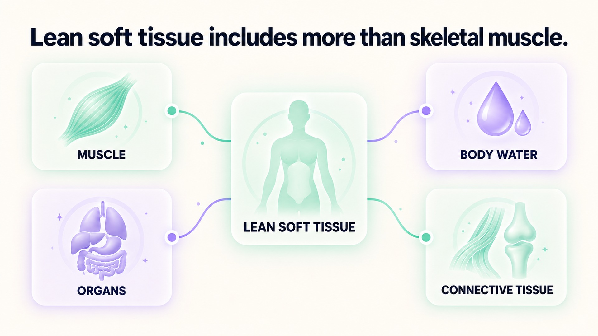
What the report does not establish
A DXA category is an estimate created by a specific imaging method and analysis. The scan does not identify the biochemical makeup of every pixel or separate each organ, tendon, and muscle on its own. The software classifies signals into the compartments its model supports. A displayed value may therefore be useful for comparing certain measurements while still leaving questions about what changed within the broader compartment.
The same caution applies when a device labels a field “muscle mass.” That label may be derived from a predictive model or a regional lean-tissue value rather than a direct measurement of skeletal muscle. If the method is not described in the report, ask the clinic what the field represents and whether it was measured, estimated, or calculated. Record that answer beside the result. Do not infer a method from the number of decimal places or from the device's marketing description.
A whole-body result and an appendicular result also answer different questions. Appendicular values refer to specified limb regions, while a total-body value includes additional areas. A height-adjusted index introduces another calculation. When reports use shortened terms such as ALM, ALST, or “lean mass,” copy the term as printed and ask which tissue and region it covers. This avoids combining terms that look similar but were defined differently.
A scan is one piece of a broader evaluation. Training records, symptoms, function, food intake, and other clinical measures answer separate questions. The scan cannot establish whether a person is strong, well nourished, or progressing in a particular exercise. If a clinical decision depends on a body-composition result, the responsible professional should explain which compartment matters and whether another method or observation is needed.
How to compare two reports
First check that both reports refer to the same person, body region, units, scan type, and analysis convention. Then compare the dates, preparation instructions, and any note about positioning or scan quality. If software or equipment changed, ask whether the clinic considers the old and new values directly comparable. A report can be technically complete while still needing an explanation before it is used as a trend.
Avoid subtracting a number from one report and calling the difference a change in muscle. If the clinic reports a change below its own expected measurement variation, it may not be possible to distinguish that difference from measurement noise. A professional can explain the site's precision, the scan's intended use, and whether a repeat at an appropriate interval would answer the question. The reader should not invent a universal “meaningful change” threshold for all machines and body regions.
Keep original reports even when a dashboard creates a simpler chart. The dashboard may round values, combine body regions, rename a field, or omit quality notes. The full report makes it possible to verify the source measurement and understand what the chart is summarizing. If the body-composition result is copied into an app, retain a note that it came from a scan and preserve the report's exact component name.
Keep the scan details with the trend
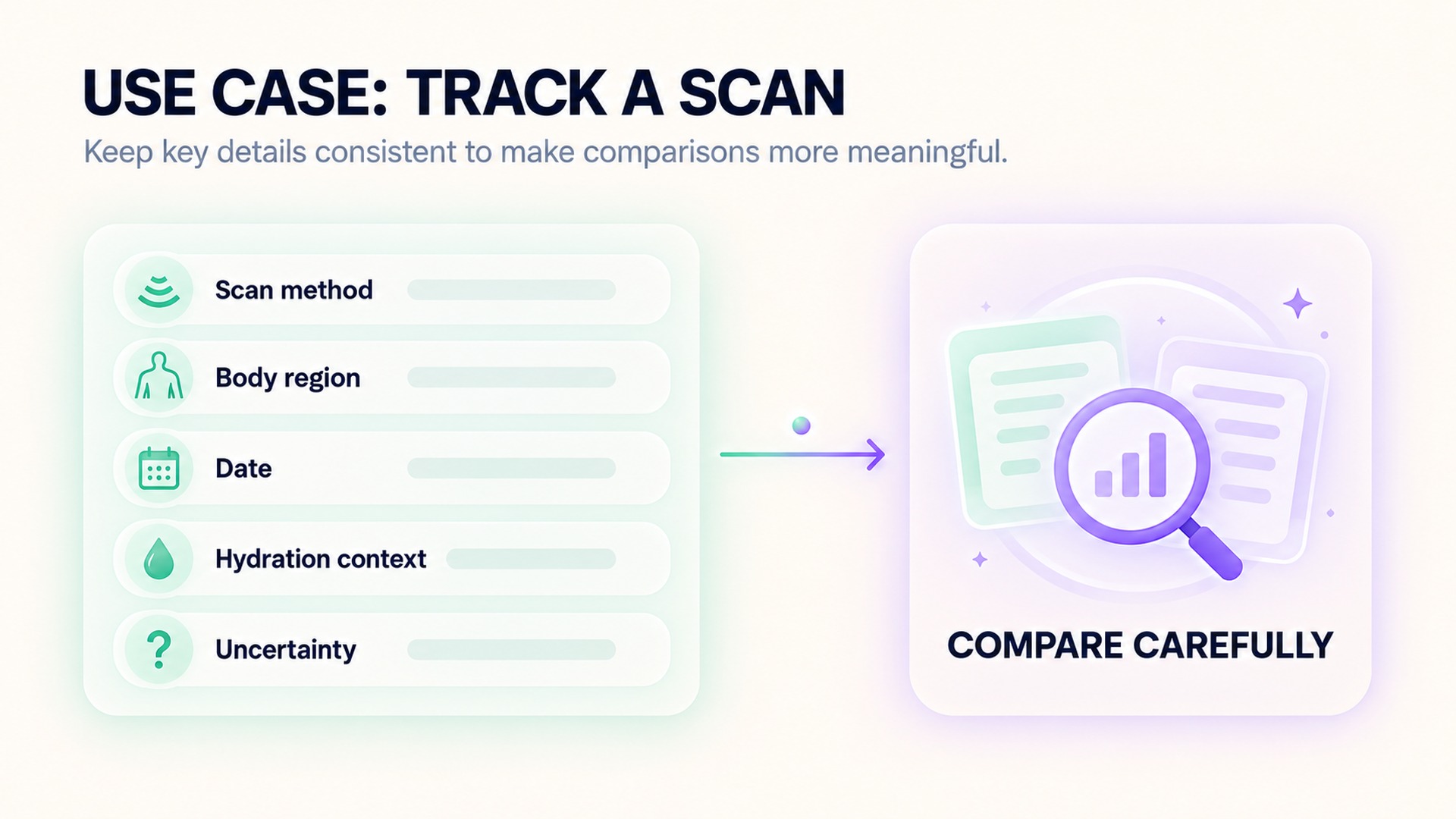
Save the complete report with scan date, equipment or method, body region, units, and the exact compartment label. If the report separates arms, legs, trunk, or other regions, keep those fields distinct from total-body results. A regional value should not be substituted for the whole-body value or combined with a result from a different region.
Record relevant conditions if the testing team provides them. Note whether the scan followed the clinic's preparation instructions, whether hydration or recent activity was flagged, and whether there were changes in equipment or software between visits. Do not try to adjust a result yourself to account for water, exercise, or another factor. The clinician or imaging professional can explain whether a comparison remains meaningful.
A short-term body-weight change and a scan compartment are different observations. A related article on why short-term weight changes do not identify body-fat change explains why a scale value cannot identify which tissue changed. A scan offers more compartment information, but it still depends on the method and does not directly measure every muscle.
Use a trend only when the reports are comparable enough to answer the question. A change in scan equipment, analysis method, body region, or report terminology may require a break in the series. Keep the original reports rather than copying one number into a generic “muscle mass” field. If a tracker uses a simplified label, mark the underlying test and exact measurement name so a later reader can see what was measured.
The scan can still be useful without being treated as an exact muscle meter. It provides a method-dependent estimate of body compartments that a care team or qualified professional can interpret alongside other information. I keep the report's wording, date, region, and conditions visible. That lets the measurement contribute to a broader assessment without turning lean soft tissue into a claim that the scan did not make.
Frequently asked questions
Does lean tissue on a scan equal skeletal muscle?
No. Lean soft tissue is a broader compartment that includes skeletal muscle and other nonfat soft tissues, including water. A DXA report does not directly weigh each skeletal muscle.
Can hydration affect a lean-tissue result?
Hydration can affect the amount of water included in lean soft tissue. Keep preparation conditions and the scan method with the report, and ask the testing team whether two results are comparable.
Should I use a scan change as a muscle-gain number?
Not by itself. The reported compartment can change for reasons other than muscle, and the scan has method-specific uncertainty. Review the full report and context with the clinician or professional interpreting it.
*This article is for informational purposes only and is not a substitute for professional medical advice, diagnosis or treatment.*
Sources:
The American Journal of Clinical Nutrition·The journal of nutrition, health & aging·American Journal of Clinical Nutrition·Obesity Pillars·European Journal of Nuclear Medicine and Molecular Imaging·The British journal of nutrition
Related articles

Urine albumin concentration needs its creatinine context
A spot urine albumin concentration changes with how diluted the sample is. The albumin-to-creatinine ratio adds context, while the full report and clinical question still guide interpretation.

A hemolyzed blood sample can complicate a potassium result
Cell damage during collection or handling can affect a measured potassium value. Keep the sample-quality comment with the report and have the result reviewed in its clinical context.

Biotin supplements belong in the record before a blood test
Biotin in supplements can affect selected laboratory assays, and the effect depends on the method used. A clear product, amount, and timing record helps the testing team decide whether an assay-specific review is needed.

A post-vaccine HRV change is not a personal immunity test
A wearable HRV change after vaccination can describe a physiological signal, but it cannot measure antibodies or personal protection. Learn why study associations, symptoms, and immune status answer different questions.

Vitamin D blood levels should not become an automatic supplement dose
Learn what a vitamin D blood test measures and why the result does not automatically set a supplement dose. See how symptoms, medicines, testing reason, and health history shape interpretation.

Blood sodium concentration is different from sodium eaten
Learn why a sodium blood test measures a concentration while a food log estimates an amount eaten. See why a blood result cannot be converted into an instruction to add salt or change intake.

Recent lifting belongs in the context of a liver enzyme result
Learn why recent lifting belongs beside liver enzyme results and why it should not be assumed to explain an abnormal test. See how AST, ALT, CK, timing, symptoms, and the rest of the panel help shape clinical interpretation.

Keep the kidney function equation beside an eGFR trend
Learn why a change in the eGFR equation can shift a reported estimate without an equivalent biological change. See what to record beside each value so a trend can be interpreted with its method and clinical context.

Creatine kinase after exercise requires symptoms and timing
Learn why recent exercise, test timing, and symptoms all matter when a creatine kinase result is reviewed. See why CK is a clinical marker rather than a training target and when unusual symptoms need prompt attention.

Normal oxygen saturation does not establish a normal hemoglobin level
A pulse-oximeter percentage estimates how much available hemoglobin is carrying oxygen, while a blood count measures hemoglobin amount. Learn why one reassuring reading cannot replace the other test or a clinical review.