Muscle and pulse oxygen sensors measure different signals
By Mr.Apps · Nov 3, 2026
Category:Training
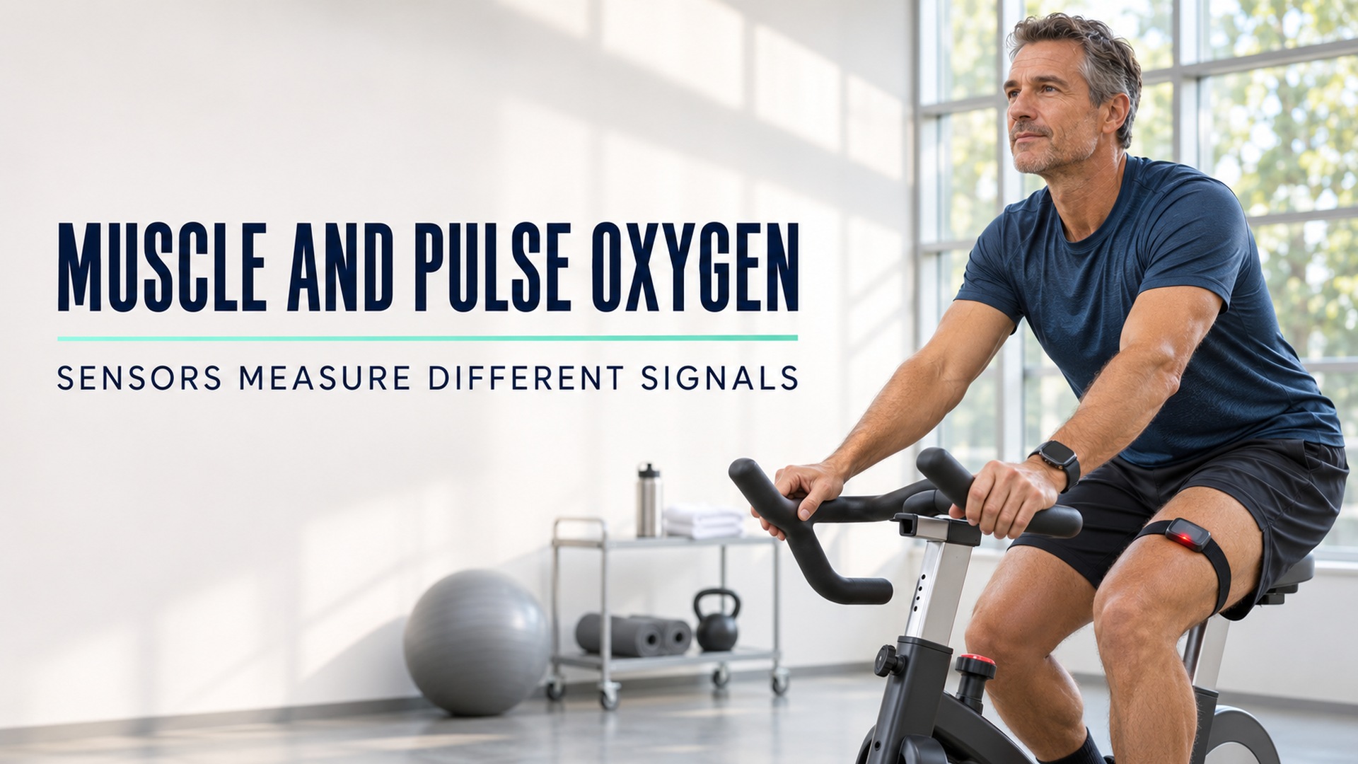
Muscle oxygen and pulse oxygen are different signals
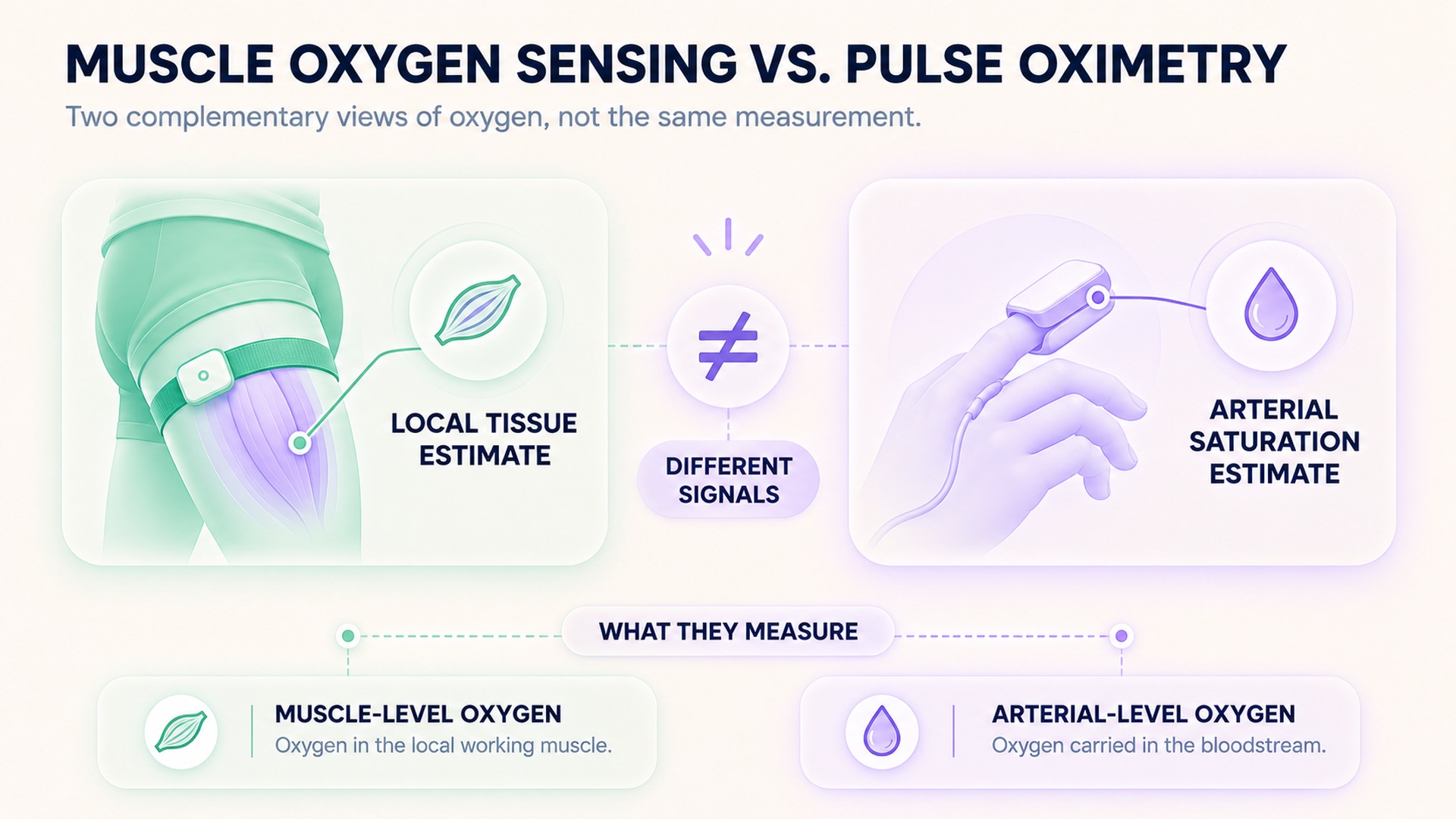
A muscle oxygen sensor and a pulse oximeter do not measure the same physiological compartment. A pulse oximeter estimates arterial oxygen saturation from the changing light signal associated with arterial blood flow at a sensor site, often a fingertip. A muscle sensor using near-infrared spectroscopy estimates a local optical signal from tissue beneath its probe. It can reflect a mixture of hemoglobin and myoglobin oxygenation and depends on device design and tissue geometry.
I keep each sensor's label and site with its value instead of comparing the percentages as one measure.
The pulse-oximeter overview describes a device that uses light to estimate blood oxygen saturation and pulse rate. By contrast, an exercise study using near-infrared spectroscopy on forearm muscle examined local hemoglobin and myoglobin oxygen desaturation under a specific protocol. The two displays can both use a percentage, but a shared percent sign does not make their values interchangeable.
What pulse oximetry estimates
A pulse oximeter shines light through or reflects light from a peripheral tissue site and analyzes how the signal changes with the pulse. Its displayed SpO₂ value is an estimate of arterial oxygen saturation. It does not directly measure oxygen throughout the body, oxygen use inside a working muscle, or the amount of oxygen delivered to each organ.
The signal can be affected by device quality, circulation, movement, skin temperature, ambient light, nail products, and other conditions. The draft medical-use device guidance proposes performance testing and labeling considerations. It is a draft for comment and is not for implementation. A consumer device may have a different intended use and review status from a prescription medical device. Follow the specific device instructions and the care plan when interpreting a reading.
This distinction also helps avoid over-reading a reassuring number. A pulse oximeter estimate is one data point and does not replace symptoms, examination, or clinical judgment. A related explanation of what a normal oxygen-saturation reading cannot establish about hemoglobin shows why the displayed value answers a limited question. The reading is not a complete measure of oxygen transport or blood health.
What a muscle near-infrared sensor samples
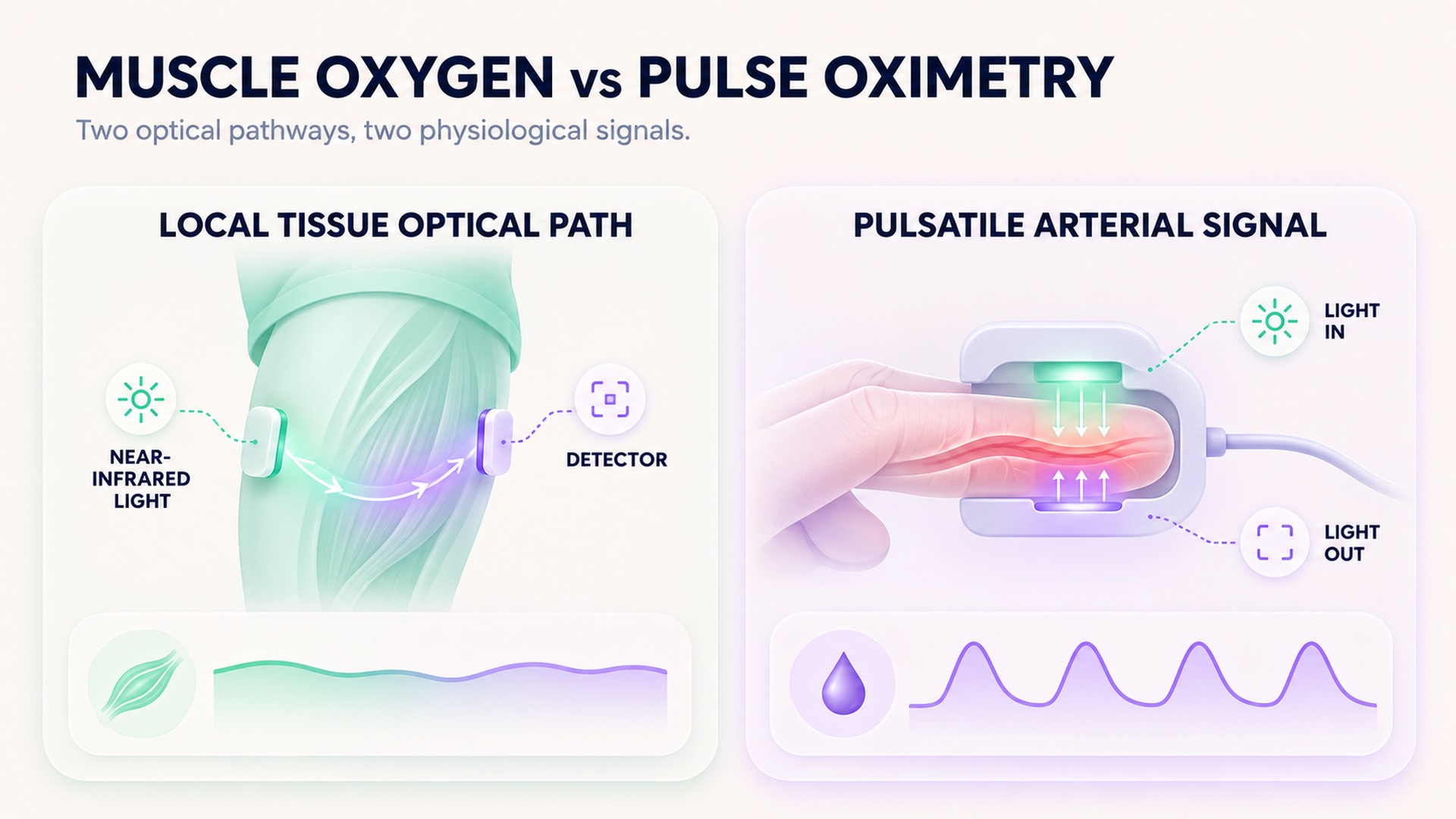
Near-infrared spectroscopy sends light into a local tissue area and measures light returning to the detector. Devices use this pattern to estimate changes in local tissue oxygenation, often over a muscle. The optical path can include skin, fat, small vessels, and muscle tissue. Depending on the device and probe placement, the output may be more muscle-weighted or reflect a broader tissue mixture.
Some muscle sensors report an oxygen-saturation value or index. That label should be read with the sensor model, placement, site, exercise condition, and method. The review of muscle oximetry in sports science discusses how near-infrared signals can reflect oxygenated hemoglobin and myoglobin in local tissue. It also notes methodological differences among devices and protocols. The review does not make one displayed percentage a universal measure across all sensors.
The device may use a label such as muscle oxygen saturation, tissue oxygen saturation, or a tissue saturation index. These names are not automatically equivalent across manufacturers. One device may apply a different algorithm, probe spacing, or correction for tissue above the muscle. Preserve the device's own field name rather than standardizing several displays into a generic “oxygen percent.” If a comparison requires a shared scale, confirm that a published validation method supports that conversion for the specific instruments.
The review of near-infrared spectroscopy in skeletal muscle describes why local oxygenation can be useful in research and exercise settings, while also noting interpretation limits. The sensor does not take a blood sample from the muscle. It estimates an optical signal through the tissue path and applies its own model. The signal may change with local blood volume, oxygen delivery, extraction, probe pressure, or superficial tissue characteristics.
Probe placement is part of the measurement. Moving a sensor to another muscle, shifting it over a different part of the same muscle, or changing how firmly it is held can alter the optical path. Skin, fat thickness, ambient light shielding, and movement can also influence the recorded signal. A local trend is easiest to interpret when the same sensor, side, placement, and exercise protocol are repeated. Those controls improve context, but they do not turn a local optical estimate into a direct reading of arterial oxygen saturation.
Why a matching percentage does not mean a matching measurement
A muscle sensor and pulse oximeter can each show a percentage while measuring different things. One reports a local tissue estimate; the other estimates arterial saturation at a peripheral pulse site. Their wavelengths, sensor geometry, signal processing, and reference assumptions can differ. Comparing the numbers directly can create a false impression that one device is “higher” or “lower” than the other on a common scale.
The denominator may not be the same either. A pulse oximeter estimates the share of hemoglobin carrying oxygen in the pulsatile arterial signal. A muscle oximeter may report a tissue saturation index based on local hemoglobin and myoglobin contributions in a mixed optical path. A similar-looking unit does not guarantee the same biological quantity or calibration.
The study analyzing hemoglobin and myoglobin components in muscle NIRS used a small group of healthy participants and a controlled forearm exercise task. It suggests that both components can contribute to the observed signal under that study's conditions. It does not establish a conversion formula from a muscle reading to fingertip SpO₂. Do not compare their values as if one could be substituted for the other.
Use the reading that matches the question
If the question concerns a local muscle response during exercise, a muscle sensor may provide a relevant research or training signal when used according to its protocol. If the question concerns an estimate of peripheral arterial oxygen saturation, a pulse oximeter is the device designed for that purpose. Neither number alone gives a full medical assessment.
The review of NIRS signal interpretation in skeletal muscle explains that tissue composition and physiological assumptions influence how a muscle optical value should be interpreted. It does not endorse using a sport sensor to diagnose low blood oxygen. Follow instructions for the specific device and seek professional guidance for clinical concerns.
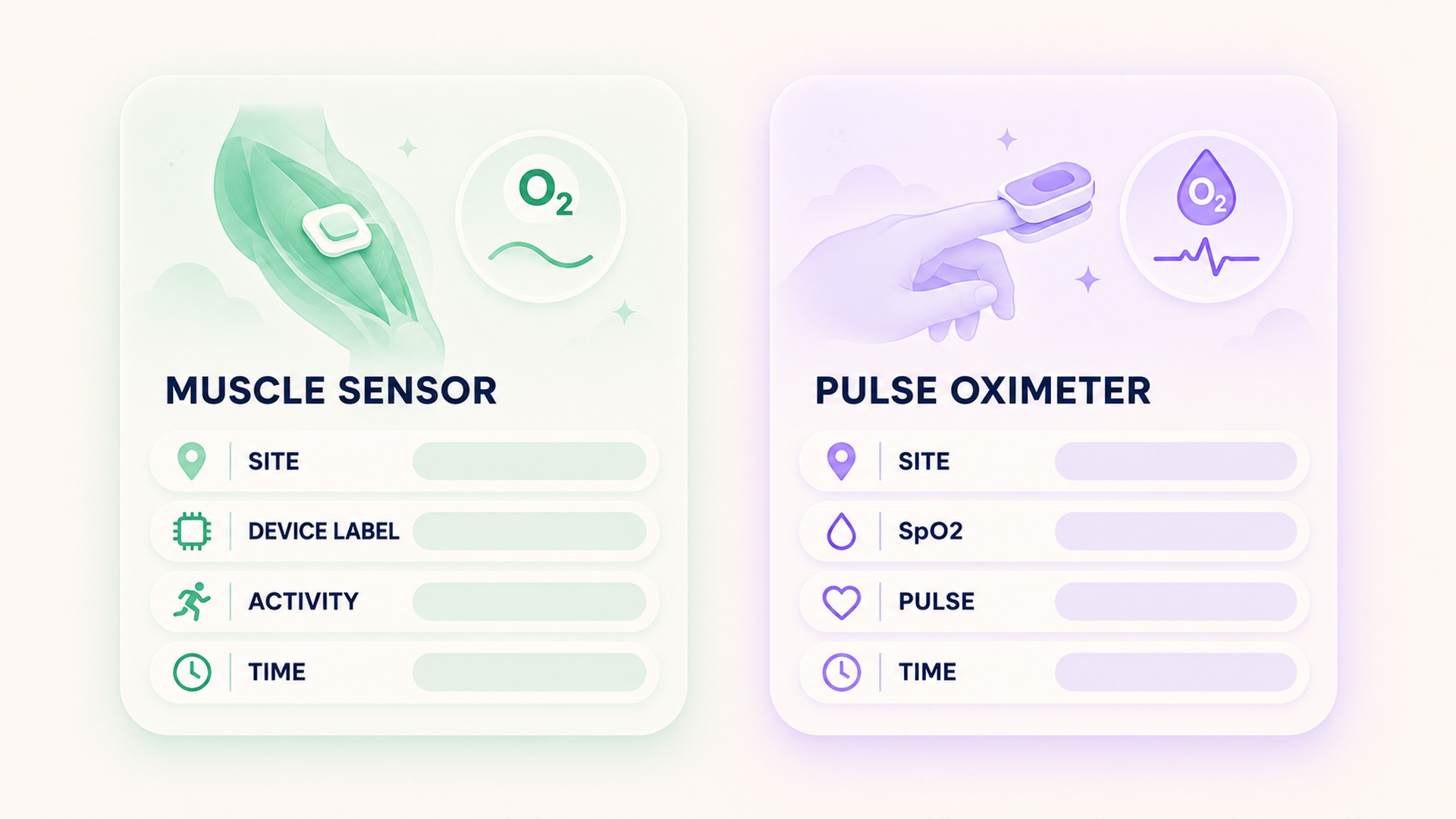
For device-to-device comparisons, retain each sensor's own label and units. Record the location, placement, activity, date, and display name. Avoid charting a pulse oxygen value and a muscle oxygen value on the same axis unless a validated method explicitly supports that comparison. A side-by-side display can be useful if the measurement types remain clearly labeled.
Keep context with the record
A useful muscle-sensor note includes the device model, sensor site, side of the body, placement method, activity phase, and whether the reading was measured at rest or during exercise. If the device reports a local saturation index rather than an arterial estimate, preserve that name exactly. Do not relabel it as SpO₂ or blood oxygen.
For a pulse oximeter record, preserve the device type, site, time, displayed SpO₂, pulse rate, and relevant symptoms or care instructions. A spot reading and a trend are different records. If the value appears inconsistent with how the person feels or with clinical guidance, follow the care plan and discuss the result with a qualified clinician. Do not wait for a muscle sensor to confirm a pulse-oximeter concern.
When reviewing an exercise file, align each value with the time and activity that produced it. A muscle sensor may show a local response while the pulse oximeter estimates arterial saturation at another site. The readings can change for different reasons and at different times. Keeping their source and measurement definitions visible prevents an appealing but unsupported interpretation.
Avoid diagnosis from a training display
A low or changing muscle oxygen value is not, by itself, a diagnosis of poor circulation, lung disease, or muscle damage. A pulse oximeter value is also an estimate, not a diagnosis. Public user guidance advises considering symptoms and seeking healthcare guidance when readings or symptoms raise concern. A person's symptoms and care plan should take priority over a training dashboard.
If someone experiences severe or worsening breathing difficulty, chest discomfort, confusion, or another urgent symptom, they should seek appropriate medical attention rather than trying to reconcile wearable numbers. This article does not set a personal oxygen threshold. The purpose of keeping the sensor type clear is to prevent one local exercise signal from being mistaken for a clinical blood-oxygen measurement.
Frequently asked questions
Does a muscle oxygen sensor measure blood oxygen like a pulse oximeter?
No. A muscle sensor estimates a local tissue optical signal, while a pulse oximeter estimates arterial oxygen saturation from a pulsatile signal at its sensor site. Their values are not interchangeable.
Can I compare the two percentages directly?
Not without a validated method that accounts for the devices and measurement definitions. Keep the sensor type, site, and display label with each value rather than plotting them as one common measure.
Can a muscle oxygen reading diagnose low blood oxygen?
No. A local muscle reading is not a diagnosis of blood oxygen status. Use a device for its stated purpose and follow clinical instructions when a health concern arises.
*This article is for informational purposes only and is not a substitute for professional medical advice, diagnosis or treatment.*
Sources:
U.S. Food and Drug Administration·U.S. Food and Drug Administration·Advances in Experimental Medicine and Biology·Sports Medicine·World Journal of Cardiology·Journal of Functional Morphology and Kinesiology
Related articles

Active and passive range of motion need separate records
Active range of motion is produced by the person, while passive range is produced by an examiner or external force. Record the method, joint, position, side, and measurement source separately because these angles are not interchangeable.
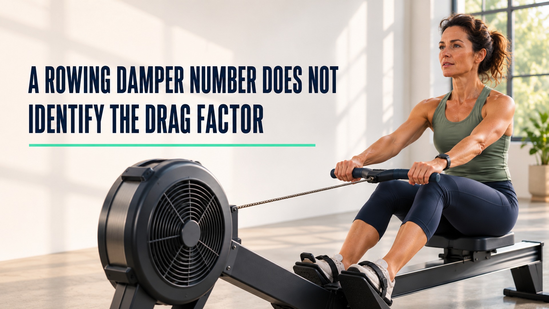
A rowing damper number does not identify the drag factor
A damper number describes an air-control setting on some rowing ergometers, while drag factor describes the flywheel response measured by a compatible monitor. Record each value with the machine model instead of treating the numbers as interchangeable.

Eccentric-only repetitions need a clear lifting-log label
An eccentric-only repetition records a lengthening action under load without an active shortening phase in the same repetition. Preserve the movement sequence, assistance, range, and tempo so its count is not confused with a full lift-and-lower cycle.

Running pace and running speed use reciprocal units
Running pace measures time per distance, while speed measures distance per time. Keep the original unit and time basis visible when converting one display into the other.
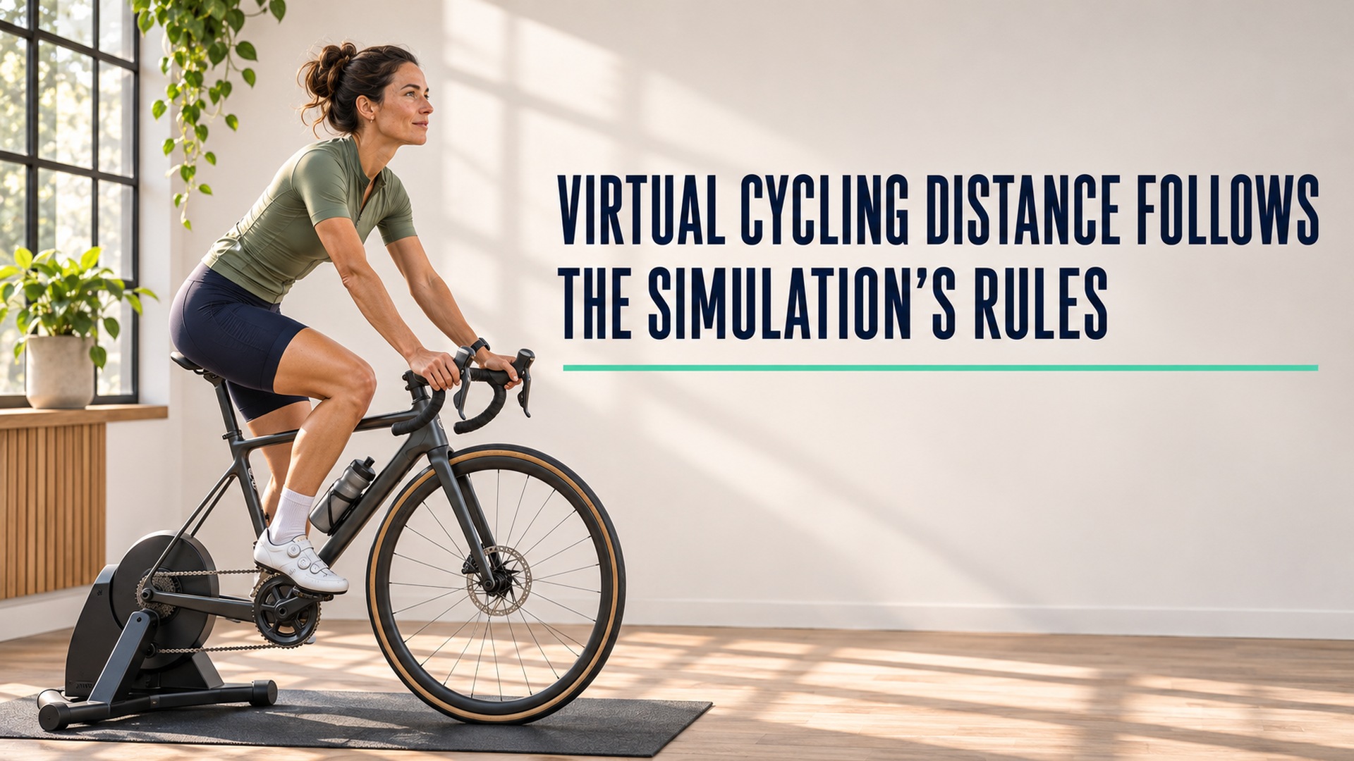
Virtual cycling distance follows the simulation's rules
A virtual cycling app calculates speed and distance from its own inputs and route model. Keep that simulated value separate from wheel-based or outdoor distance when comparing activity records.

Single-sided cycling power relies on a symmetry assumption
A single-sided power meter measures one leg and may double that value to estimate total cycling power. Preserve which side and device produced the number so an estimate is not mistaken for a bilateral measurement.
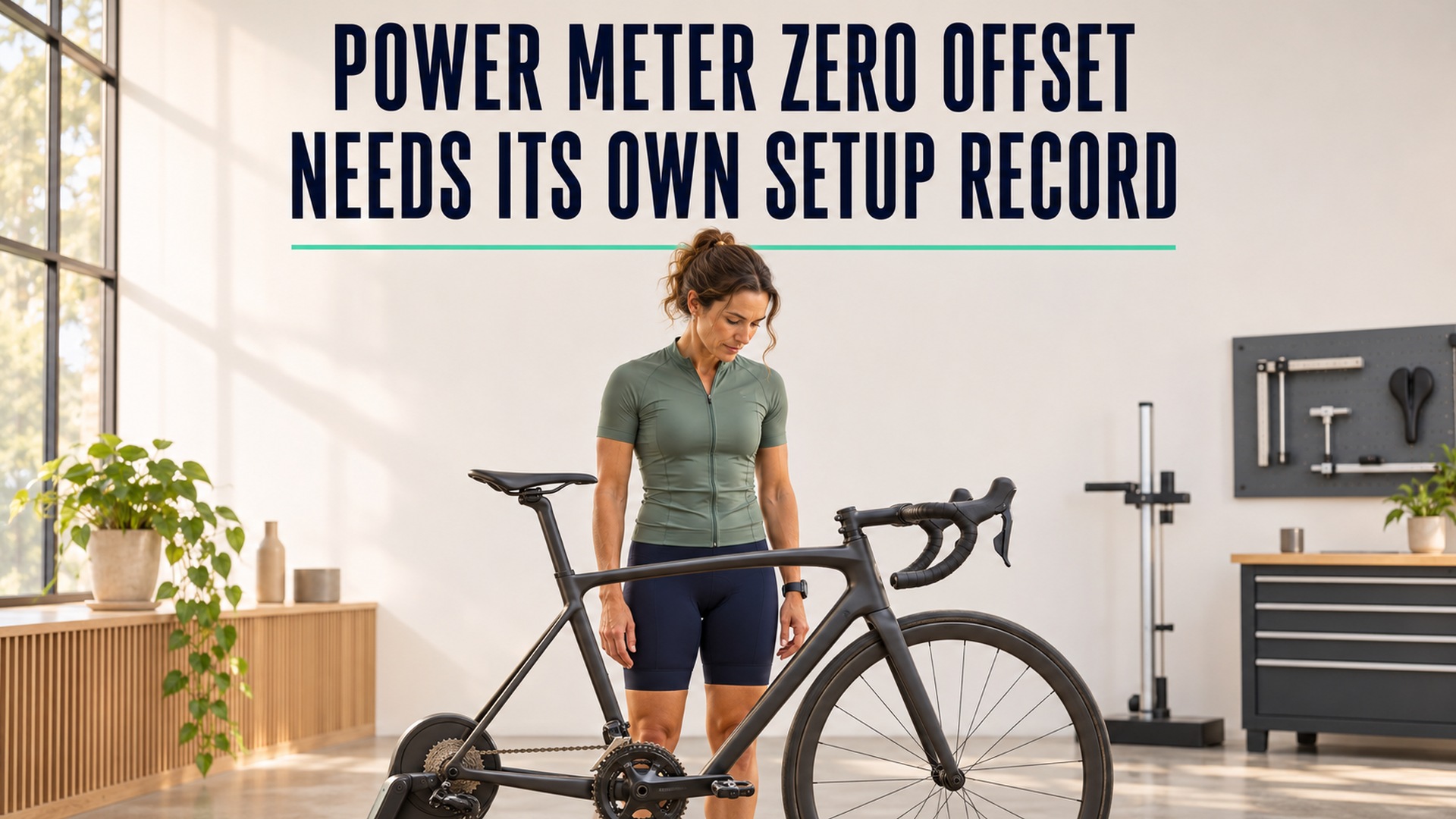
Power meter zero offset needs its own setup record
A power meter zero offset records the sensor's unloaded baseline at a particular time. Keep the meter identity, procedure, conditions, and returned status separate from training targets and workout power values.

Handrail support changes the context of a treadmill session
Treadmill handrail use can change the movement task even when speed and incline stay constant. Record whether support was light, intermittent, or sustained so session comparisons do not assume the machine measured details it missed.
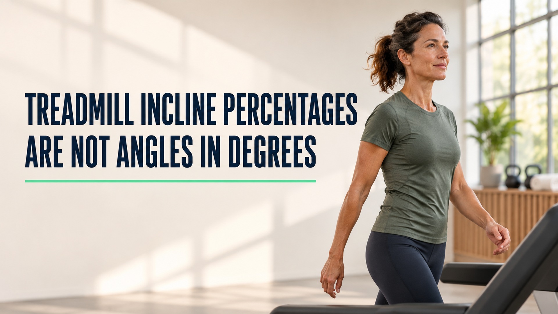
Treadmill incline percentages are not angles in degrees
Treadmill grade percentages and angles in degrees describe the same slope through different relationships. Preserve the machine's displayed unit and source so a workout setting is not mistaken for a measured angle.